Introduction
A parasite can be broadly defined as an organism that lives on or in another species, the "host", and obtains its nourishment from that species. It is easy to exaggerate the dangers, both real and imagined, of these parasites. There is no reason why fish caught by the California marine angler or bought at the market cannot be fully enjoyed even if they had once contained "guests". Parasitism is very common in nature and should not be viewed with distress. Among all the parasites found in California marine fishes, few appear to cause damage to the fish and only one, a larval roundworm, is cause for human concern. As will be seen, even this parasite is rendered harmless if the seafood is properly prepared or the parasite removed.
Tumors
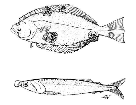 Examples of fish with tumors.
Description: Tumors and warty growths on fish can be the result of injury, pollution, bacteria, viruses, and parasites. The cause of many tumors is unknown. Although they may be aesthetically offensive to some consumers, they are not infective to humans.
Treatment: Remove tumors and handle fish as usual.
Protozoa
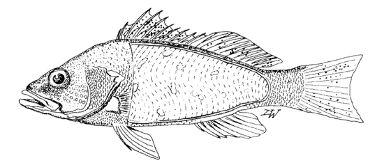 Example of a fish with protozoan infections.
Common hosts: Rockfish, herring, flatfish, and salmon.
Habitat: Skin, fins, gills, flesh, and internal structures.
Description: Protozoan infections may appear as small spots or may cover large areas of the fish. The infected area may consist of thousands of microscopic mature protozoa or spores. Although they may be unsightly, they pose no danger to humans.
Treatment: Remove parasites and handle fish as usual.
Larval Roundworms
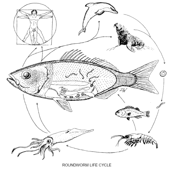 Roundworm Life Cycle.
Common hosts: Many marine fishes
Habitat: Flesh, surface of intestine and liver, and body cavity.
Description: It is easy to exaggerate the hazards, both real and imagined, of larval roundworms in the flesh of seafood. There is no reason why those who enjoy raw fish dishes, such as sashimi and seviche, cannot safely eat it if they take a few simple precautions.
Larval roundworms, or nematodes, are found in many species of marine fish. While cleaning the catch, the angler may find them coiled on the surface of the intestine, on the liver, in the body cavity, and embedded in the flesh. The larval roundworms are between 1/2 and 3/4 inches in length. When coiled they sometimes resemble a ball of yarn or a watch spring. These worms are known as "anisakids". Some live as adults in such marine mammals as seals, porpoises, whales and dolphins. The eggs enter the seawater with the feces of these mammals. They are eaten by small crustaceans such as copepods, which in turn are eaten by a fish or a squid. If this infected host is consumed by the proper marine mammal, the larvae will mature into adults. Herein lies the problem. Humans are also mammals. When we eat seafood, such as fish and squid, which contain live, infective larvae, the worms can become active in their new hosts and try to burrow into the stomach lining. This can cause lesions or growths on the stomach walls. This disease is called "anisakiasis", after the worms, and is also known as "cod-worm" of "herring-worm" disease. Some fish have lots of worms because the larvae are transferred when one fish eats another.
As mentioned above, with a minimal amount of care these worms need not be a problem. Thoroughly cooking the fish will kill the larvae. A careful examination of the flesh should be made before serving raw dishes. This can easily be done by slicing the flesh thin enough so any larvae can be seen and removed. When the flesh is held up to a light, the worms will appear as shadows. This process is called "candling". Another approach is to freeze the fish at -4° F (-20° C) for 24-60 hours. Some fish recipes, such as seviche, call for an acidic or brine-based marinade. These are not strong enough to kill the larvae. Similarly, there is a popular Norwegian recipe for "cured fish" (Gravlax) in which flesh is only slightly salted and sugared for a couple of days and then eaten with vinegar. This process, like the marinades, is not adequate to kill the larvae. Women in eastern Europe traditionally made their own gefilte fish. Those who sampled the fish before it was properly cured sometimes acquired these larvae.
Treatment: Here are a few suggestions for dealing with roundworm larvae:
- Try to clean your catch as soon as is convenient. The larvae that are coiled on the intestines may begin to move into the flesh within hours after the fish is caught.
- Cook or smoke thoroughly.
- Examine and remove all the larvae from the flesh if it is to be eaten raw or freeze it for 24-60 hours at -4° F (-20° C).
- Don't count on the pickling, curing processes, or home refrigerators (most of which cannot attain the cold temperature required) to kill the larvae.
Flukes and Roundworms
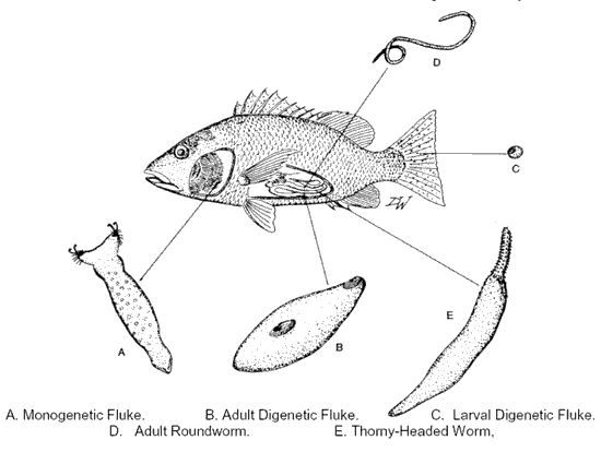 (A) Monogenetic Fluke (B) Adult Digenetic Fluke (C)Larval Digenetic Fluke (D) Adult Roundworm (E) Thorny-Headed Worm.
Common hosts: Many marine fishes.
Habitat: Skin, fins, gills, flesh, and internal structures.
Description: The above parasites are common in fish, but usually are not noticed by anglers. The monogenetic flukes (A) are often found on the gill filaments and in the gill cavity, on the eyes, and in the mouth. They are flat and may vary in shape from round to elongate. They remain attached to the host by means of small clamps or hooks. When present in large numbers, some species damage gill filaments. These flukes are called "monogenetic" because their life cycle is direct; that is, between hatching from the egg and settling on their final host, they do not live on an intermediate host.
If the gut is opened and examined closely, the angler will often be rewarded by finding several interesting types of worms. Unlike the monogenetic flukes, the digenetic flukes (B) must pass through several other hosts, including a snail, before returning to the final host to become adults. Larval digenetic flukes (C) are often seen as small cysts on the interior of fish, or on the fins and gills or in the eyes. These flukes may vary in size and shape. They may be flattened or sausage-shaped, rounded or elongate. They may vary from 1/16 inch to several inches in length. One species found in sharks is the size and shape of a cigarette. Adult roundworms (D) may be red or whitish and may reach an inch or so in length. During their life cycle they may have passed through zooplankton, small crustaceans, and/or small fish before entering this suitable, final host. Other species give live birth to their young in the flesh of fish. The thorny-headed worms (E) are a little stubbier than the roundworms. They get their name from the thorn-like hooks on their proboscis. These worms may cause damage to the inside of the fish's intestine. Larval stages are usually found attached to the gut.
Treatment: Remove parasites and handle fish as usual.
Leeches
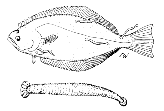 (1) Example of a fish with leeches (2) Closeup of a leech.
Common hosts: Sharks, skates, flatfish, cod, salmon, rockfish, and cabezon.
Habitat: Exterior surfaces, fins, anus, gill cavities, and spiracle valves.
Description: Leeches are generally elongate, flat, and have suckers on each end. The smalI sucker surrounds the mouth. They can often be seen "inch-worming" their way across the host's body surface. On rough skinned hosts, such as skates and sharks, leeches are found most often on the host's soft stomach or in the spiracle valves.
Typically, leeches feed by making a series of small cuts on the fish's skin and then ingesting the blood. An anticoagulant in the mouth of the leech prevents the blood from clotting. To further aid feeding, leeches inject histamine into the host's blood vessels. This causes the vessels to dilate, thus increasing the blood flow. Some species need to feed as often as once per week, while others feed only every 6 months or so. Some can carry blood parasites from fish to fish in the same way that mosquitoes carry malaria between humans. Many fish leeches leave their hosts to lay their egg-like cocoons. A single young leech emerges from each cocoon.
Until relatively recently, millions of medicinal leeches were used annually in the practice of bloodletting to treat a variety of human ailments.
Treatment: Remove parasites and handle fish as usual.
Tapeworms (Adults)
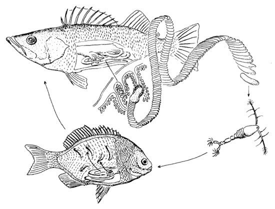 Life cycle of a tapeworm.
Common hosts: Rockfish, rays, sharks, bass, perch, salmon, and tuna.
Habitat: Stomach and intestine.
Description: Fish tapeworms, also called broad tapeworms, may range in size from an inch to more than a foot in length. They are most often seen crawling from the anus or when the gut is accidentally cut while cleaning the catch.
The scolex (head) is used for attachment. The tapeworm has no mouth and so it absorbs all its nutrients directly through its body wall. The tapeworms' body grows by producing small segments (called proglottids) behind the scolex, hence the name "tapeworm". The tapeworm's body is a series of reproductive units. The proglottids closest to the scolex are sexually immature. Tapeworms can self-fertilize and as the proglottid develops the eggs inside are fertilized. By the time the proglottid reaches the end of the worm it is full of ripe eggs. The eggs leave the fish or shark with its feces. Depending upon the type of tapeworm, the egg may be eaten by a microcrustacean, such as a copepod, or a small larva may hatch from the egg, swim freely and then be eaten by a small crustacean. The larval tapeworm undergoes development in the crustacean. When this infected host is in turn eaten by a fish a second type of larval stage develops. This larva will remain embedded in the flesh or viscera until it is eaten by the proper species of fish or shark. Here it will mature and begin the life cycle once again.
Treatment: Remove parasites and handle fish as usual.
Lesions in Striped Bass from San Francisco Bay
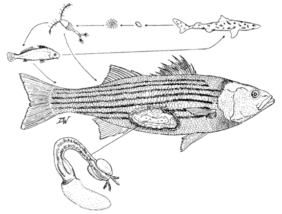 The life cycle of the larval tapeworm: Lacistorhynchus tenuis.
For years fishermen have noticed red marks and open lesions on the right sides of striped bass from San Francisco Bay. These sores are caused by a larval tapeworm (Lacistorhynchus tenuis). The adult tapeworm lives in the intestines of sharks and rays. The tapeworms' eggs pass out with the hosts' feces. A free swimming larva hatches and is eaten by a small crustacean. The infected crustacean is in turn consumed by a fish and the larva, about the size of a grain of rice, embeds itself in the viscera or flesh of the new host. The larva will remain there until it is eaten by the correct species of shark or ray and then it will develop into an adult. However, when this larval stage is eaten by striped bass from San Francisco Bay, the larvae are killed and encapsulated with fibrous tissue. If there are lots of larvae the fibrous tissue will form a "raft" in the viscera. The striper can also become infected if it eats other fish that contain the larvae. Hence, the number of larvae consumed can build up very quickly. The reddening or "strawberry" mark seen by the fisherman is the inflammation caused when these "rafts" touch the inside of the body wall. The striped bass further reacts to the "raft" by encapsulating it with its own muscle tissue. As a result an open lesion is produced on the outside of the fish. The lesions are usually on the right side of the fish because of the arrangement of the internal organs. Interestingly, this larval tapeworm is found world wide in over 100 species of fish, including east coast striped bass and other species of San Francisco Bay fish, with no reported pathology. This type of reaction is unique to San Francisco Bay striped bass.
Treatment: Remove parasites and handle fish as usual.
Isopods
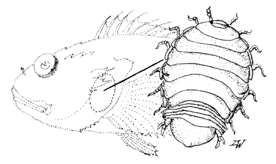 An isopod.
Common hosts: Rockfish, surfperch, and especially bottom fish such as lingcod, cabezon and flatfish.
Habitat: Gill chambers and mouth.
Description: These crustaceans, distant relatives of the garden pillbugs, range from 1/8" to more than an inch long. Their body is whitish and they have two small black eyes. The main part of their body is divided into seven segments, each of which has a leg attached on each side. The legs are armed with sharp sickle-shaped tips which help the isopod attach to its host. These sharp appendages also are responsible for many of the "bites" suffered by unwary collectors. The next five smaller, partly fused segments are the abdomen, followed by the tail.
In most isopods the sexes are separate and relatively easy to tell apart. The males tend to be smaller than the females and have no eggs under their abdomen. Some isopods display a phenomenon known as protandrous hermaphroditism (being a male when young and then developing into a female). This process begins when the young parasite, then a male, attaches to the host. In time the male will become a female, develop a brood pouch and produce eggs. If however, there is a female already present on the host when the young male first arrives, the male's development into a female may be delayed or it may remain a male as long as the female stays on that host. In Icelandic sagas these parasites are called "Peters' Stone". A dried isopod was worn as a charm or powdered and taken as a remedy. Isopods living in the gill chamber may graze on the gill filaments or cause skin lesions.
Treatment: Remove isopods and handle fish as usual.
Copepods
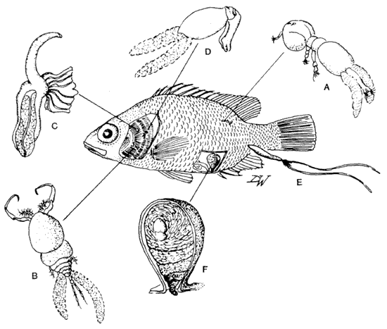 Types of copepods.
Common hosts: Most marine fishes.
Habitat: Exterior surfaces, mouth, gills, body cavity, and eyes.
Description: Copepods, like isopods, are crustaceans. These parasites vary in size, shape and position on or in fishes. Copepods can be grouped into roughly three categories: 1) those that crawl freely over their hosts and move from fish to fish, 2) those that are less motile and show some structural reduction, and 3) those that are permanently fixed to a host and show much structural reduction. Some copepods (A) crawl freely over the surface and can move from fish to fish. These species are about 1/4" long. Their flattened shape and sharply pointed appendages allow them to move around without much water resistance while holding tightly to their hosts. This type of copepod (there are over 400 species) resembles some of their free-living relatives and are sometimes referred to as "sea lice."
Some copepods attach to the lining of the mouth and gill cavities and to gill filaments. Compared to the copepods that crawl over the body, this second group is less mobile and may have reduced appendages. One type (B) has large sickle-shaped attachment claws. This species can severely damage the gill filaments. Some species can wrap their ribbon-like appendages around a gill filament (C) and feed on nutrients sucked from the gills. Other copepods (D) may be found on the gills or attached to the lining of the mouth or gill cavity. This group contains over 1000 species.
A third group of copepods has completely traded mobility for security. They may be found burrowed into the flesh of the fish's body (E) or may penetrate the eye and embed in the retina. Some of the burrowers will penetrate blood vessels or even the heart. The younger stages make a hole in the skin, insert their button or horn-like head, and when the skin heals around the wound, the copepod is permanently attached. When the copepod dies the head may form a cyst in the flesh. These copepods have undergone such radical modifications that there is little left besides their digestive and reproductive tracts. One of the most extreme examples of this is the copepod Sarcotaces. It is found in the body cavity near the anus. Rockfish are a common host. The male and female copepods are surrounded by a sac made from intestinal tissue (F). The large female appears to be little more than a pear-shaped bag covered with wart-like growths. The males are much smaller than the female and shaped like an arrow. If the sac is cut while cleaning or filleting the fish, a black, inky fluid will leak out. This is degenerate fish blood which filled the body cavity of the copepod.
In addition to modifications of their morphology, copepods have adapted to the parasitic way of life in several other interesting ways. The problem of finding and keeping a mate has been solved by one species by having a female that is 12,000 times larger than the male. Since the male's only function is to fertilize the female (like a drone in a beehive), it is advantageous to have a dwarfed male that attaches for life to the first female it finds. The male receives nourishment from the female and in turn, serves as a reproductive organ for the female. In other copepod species a permanent mate is not as important. One copulation fertilizes a female for life. Even with such reproductive efforts as those described above, the chances of any one copepod egg completing its life cycle is extremely small. To improve these chances, one species (B) releases more than 100,000,000 eggs per year. In general, parasites that have complex life cycles produce many eggs. Obviously, the more complex the life cycle, the more hazards the larval forms will encounter and the greater the number of offspring that must be produced to balance the larval loss.
Treatment: Remove parasites and handle fish as usual.
References
Hilderbrand, K. S., R. J. Price, and R. E. Olson. 1994. Parasites in marine fishes: questions and answers for seafood retailers. http://seagrant.oregonstate.edu/sgpubs/onlinepubs/g03015.pdf. [last accessed 22 June 2011].
Kabata, Z. 1970. Diseases of fishes. Book 1. Crustacea as enemies of fishes. T. F. H. Publ. Jersey City, New Jersey. 171 p.
Mayes, M. A. 1969. What's bugging the fish? An angler's guide to fish diseases and parasites. Nebraska Game & Parks Comm. 8 p.
Myers, B. J. 1975. The nematodes that cause anisakiasis. J. Milk Food Techn., (38):774-782.
Olson, R. E. 1986. Marine fish parasites of public health importance. In: Kramer, D. E., and J. Liston, ed., Seafood Quality Determination. Elsevier Science Publishers, Amsterdam: 339-355.
Oshima, T. 1972. Anisakis and anisakiasis in Japan and adjacent area(s). In Morishita, K., Komiya, Y., and Mastsubayashi, H., ed., Progress in medical parasitology in Japan, 4: Meguro Parasitological Museum, Tokyo: 303-393.
_____,1976. Research then and now on the Anisakidae nematodes. Trans. Am. Micro. Soc., (95):137-142.
Schell, S. C. 1970. How to know the trematodes. Wm. C. Brown Co. Publish. Dubuque, Iowa. 355 p.
Schmidt, G. 0. 1970. How to know the tapeworms. Wm. C. Brown Co. Publish. Dubuque, Iowa. 266 p.
Schultz, G. A. 1969. How to know the marine isopod crustaceans. Wm. C. Brown Co. Publish. Dubuque, Iowa. 359 p.
Sindermann, C. J. 1970. Principal diseases of marine fish and shellfish. Academic Press. New York and London. 369 p.
Yamaguti, S. 1963. Parasitic copepoda and branchiura of fishes. Interscience Publish. New York, London, and Sydney. 1104 p.
From Common Parasites of California Marine Fish, Leaflet No. 10, by Mike Moser and Milton Love, published by the State of California, Department of Fish and Game. This publication is no longer in print.